The concept of Picture Archiving and Communication System (PACS) lives on as a cornerstone for the various advancements to the digital imaging domain, contributing to the sleek acquisition, distribution, visualization, and automated analysis of medical images. Moreover, medical professionals worldwide still rely on this kind of digital platform as part of their clinical workflow, which in many cases is shaped by what their PACS has to offer.
PACScenter is a living product which continues to receive new features, thanks to the work of our engineers and the feedback collected from our clients. With the second month of 2024 just starting, we will showcase the latest features in PACScenter that are readily available for new and existing users.
Visual Overlays
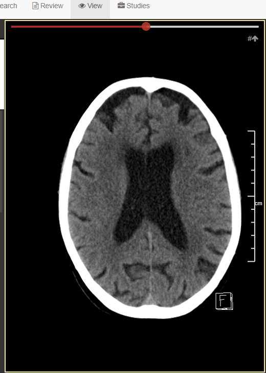
There may often be a need to superimpose certain shapes or masks on top of an image, representing a segmentation of a particular finding. In some scenarios, additional information may already be available, but only in the DICOM overlay data. PACScenter today will show DICOM overlay data on top of the images if they have one, and they can be toggled on and off.
An example of an image with a DICOM overlay to present plane reference hints.

This is just one of the ways where overlays make a difference. They also can uniformise volume and surface mask presentation alongside the existing annotation system, to streamline the integration of machine learning segmentation models.
DICOM Structured Report Visualization
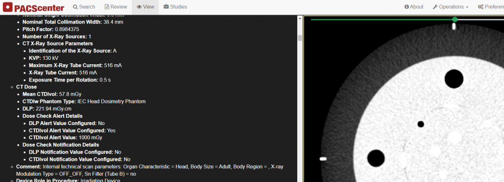
Structured reports (SRs) as established by the DICOM standard are highly versatile pieces of information, empowering systems to create machine-readable documents with a rich and well defined structure. As a noteworthy example, many computer assisted detection (CAD) systems already print their findings and early conclusions as DICOM SR. Even outside the booming medical AI industry, some devices produce DICOM SR for extended acquisition metrics and radiation dose reports.
The latest PACScenter is aware of DICOM SR files in a study, and allows you to inspect their contents alongside the images in that study. A new textual view presents the information in the file through a comprehensive translation of the DICOM data into hierarchical markup, and concept values are automatically translated to their human-readable descriptions. If the report contains references to images in the study, these references will appear in the report as clickable links that open the image when clicked.

In addition to viewing these files as a whole, it is now possible to retrieve a particular piece of information from an SR file, specified using an XPath-like syntax, and map it into a label in the label profile, making it visible alongside the other details in the label overlay. This makes it possible for doctors to keep the current workflow of inspecting meta-data around the images under observation.
Peek or toggle between the current and previous mammography scans
We had a request for a quick and easy way to switch between the current and previous scans in mammography, as it is particularly helpful to highlight the differences between the last acquisition.
As of the latest version, it is now possible to assign a shortcut key to the new study peeking tool. Then the key is held down, the viewer will show the corresponding images from the previous or current exam, per the same layout. For example, when viewing the latest mammography acquisition, a medio-lateral oblique (MLO) scan of the left breast on the lower-right view will be replaced by the MLO scan of the left breast from the previous exam on the lower-right, and so on. Releasing the shortcut key will revert the views back to the other study.
Navigation direction on breast tomosynthesis
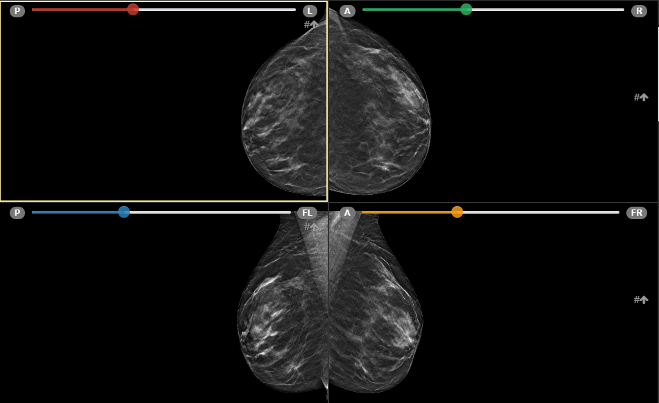
Breast tomosynthesis is a particularly demanding modality, and sometimes the direction of navigation over the multiple slices of acquisition is not entirely clear. To help clear eventual doubts, the viewer will now show patient-oriented direction letters in both ends of the navigation slider when a tomosynthesis stack is shown.

Region-based pixel data anonymization
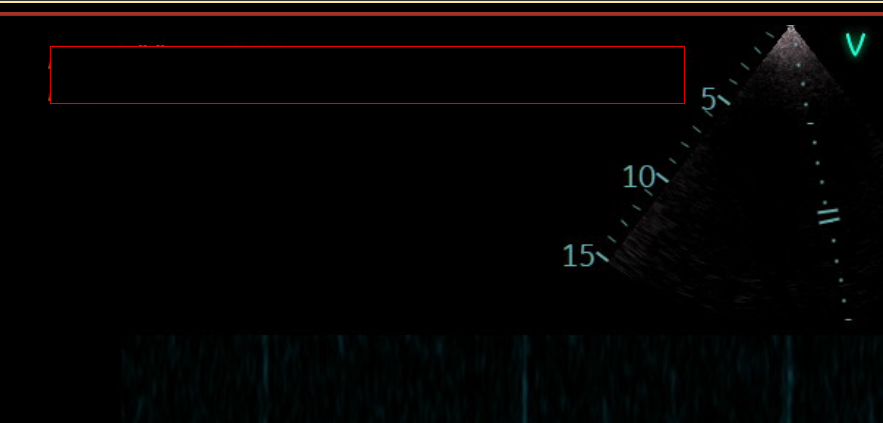
For a while PACScenter has offered an automatic approach to pixel data cleaning when downloading anonymized DICOM studies. Now, the user can also annotate the regions to remove from the image, using a new Region tool in the viewer itself. This can be useful in cases where the private information burned in the image is always in the same positions (which is often the case in ultrasound scans), or when an automatic image deidentification algorithm is not available.

Better disk caching support and use of modern browser capabilities
In past iterations of PACScenter, new users on Firefox would be shown a notice suggesting them to use a modern version of Chrome. This was primarily due to the fact that disk-based image caching was only available on Chromium-based browsers. With the latest versions, PACScenter features a disk-backed cache implementation depending exclusively on established Web APIs, thus working on Firefox, Safari, and many other browsers. Despite the usually controlled environment that is a medical imaging workstation, it is our intention for PACScenter to work well and provide a fluid experience on a standard modern Web browser.
We are also keen on taking advantage of browser capabilities when we believe that they are sufficiently widespread, especially to users who keep their browsers up to date. For the first time in PACScenter, we were able to confidently deliver WebAssembly modules alongside the main web application, which made it possible to implement DICOM structured report parsing directly in the browser.
If you would like to know more or request a demo with us, please feel free to reach out to info@bmd-software.com.
