BMD Software remains committed to developing the next generation of digital pathology solutions. BMD Software’s PathoBox is a 100% web-based, zero-footprint solution designed to bridge the gap between microscopic analysis and high-performance clinical data management.
Zero-Footprint interoperability: The DICOMweb Advantage
As a high-performance Image Manager and Image Display, PathoBox defines the new standard for interoperability. While legacy systems remain “closed”, our DICOMweb-native architecture ensures seamless, zero-footprint integration across complex hospital ecosystems. PathoBox extends beyond basic image viewing to support a more comprehensive diagnostic workflow. By integrating macroscopic and microscopic data, it provides pathologists with the full clinical context needed for complex cases. Particularly, key features include
Enterprise-Scale WSI: Native support for Whole Slide Image (WSI) concatenation (Fig.1).
Integrated Context: Full macroscopic image visualization alongside microscopic slides (Fig. 2).
Precision Control: Manual scale assignment for exact diagnostic context.
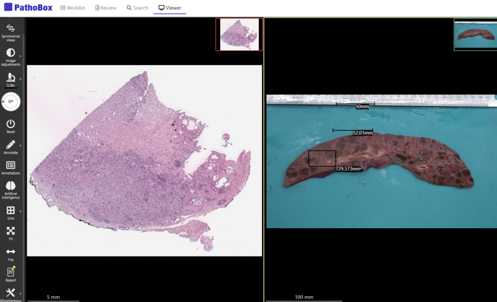
Fig. 1 – Integrated macroscopic visualization: A side-by-side view of macroscopic (gross) tissue and microscopic slides, featuring manual scale assignment for better context.
Interoperability excellence: DICOM Success
One of the highlights last year for BMD Software was participating in the DICOM WG-26 Connectathon as both an Image Manager and an Image Display. During the event, PathoBox’s archiving and visualization interfaces were tested by other participants.
PathoBox successfully displayed images from most participating modalities and image managers. Other image display systems were also able to retrieve and visualize images stored in PathoBox. As shown through previous projects, BMD Software has consistently demonstrated a strong commitment to the DICOM standard. Across the industry, professionals widely recognize interoperability as a key factor in achieving an effective digital pathology workflow.
AI Innovation: Lung Cancer Analysis
Within the scope of the HfPT 5.3.2 PRR Project, in collaboration with the Faculty of Medicine of Coimbra and CCG Research Institute, 170 lung cancer slides were uploaded to PathoBox and used to develop two new algorithms:
- Lung adenocarcinoma segmentation (Fig. 2)
- Epithelial cell segmentation (Fig. 3)
The algorithms were developed using an active learning loop, facilitated by PathoBox’s API. Expert pathologists annotated a small subset of images directly on PathoBox, which were used to train a baseline version of the algorithm. The algorithm then annotated new images, which pathologists reviewed. PathoBox designed new interfaces and frameworks to support both research and clinical environments, positioning it as a flexible and versatile solution.
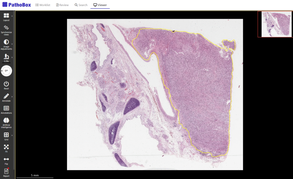
Fig. 2 – Lung Adenocarcinoma Segmentation: The AI algorithm defines tumor boundaries with a yellow outline, helping pathologists quickly identify areas of interest.
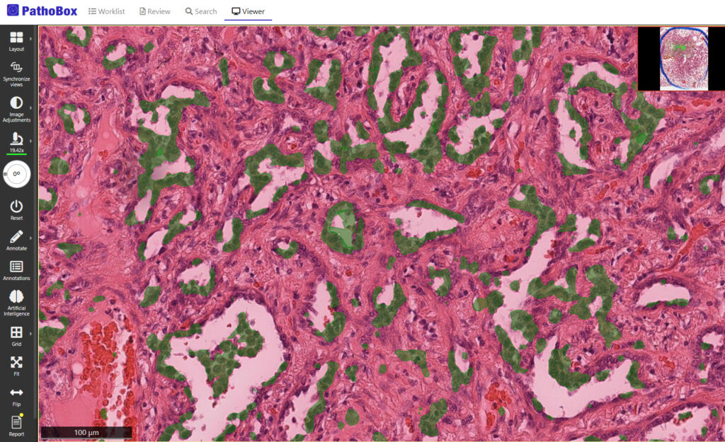
Fig. 3 – Epithelial Cell Segmentation: The PathoBox algorithm automatically identifies and highlights epithelial cells in green to assist in detailed tissue analysis.
Analyzing feedback from pathologists at SPAP
We showcased the algorithms and new features at the 20th National Congress of Pathological Anatomy in Portugal (SPAP). To better understand how users perceive PathoBox, we conducted a user feedback survey after pathologists interacted with the software. The results were highly encouraging, as demonstrated in Figure 4: 96% of respondents indicated that PathoBox meets their needs well or very well, with 56% reporting “very well” and 40% “well”, while only 4% expressed a neutral opinion.
Therefore, these results confirm that the platform is effectively addressing real-world requirements and provide strong motivation to continue investing in usability, interoperability, and advanced image analysis capabilities.
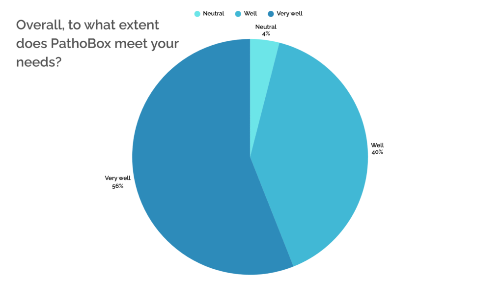
Fig. 4 – Satisfaction survey: PathoBox received high marks, with 96% of users reporting that the platform meets their needs “Well” or “Very Well.”
As we move forward into 2026, BMD team will continue to enhance PathoBox’s DICOMweb integration. The team will also further develop its Image Analysis framework to support a broader range of tools and use cases.
Discover more about PathoBox in other related news:
- PathoBox – Advancing Healthcare in Anatomical Pathology
- BMD showcases PathoBox at IHTC 2025 in Porto
- Showcasing Pathobox at SPAP National Congress

This digital pathology solution is a project in the context of Health from Portugal – “Agenda Mobilizadora para a Inovação Empresarial” (grant agreement No C644937233-00000047).